
Figure 1.
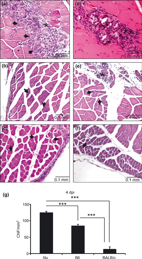
Histological analysis of Triceps brachii stained by H–E at 4 (a, d), 8 (b, e), and 12 (c, f) days after intramuscular Bp injection in C57 (a, b, c) and BALB/c (d, e, f) mice. Arrows show regenerating myofibres and asterisks, the inflammatory infiltrate. (g) quantitative analysis of centrally nucleated fibres (CNFs) in the lesion area. Nu = BALB/cnu/nu, B6 = C57BL/6. Results are expressed as mean (±SD). Three animals per group were included. ***P < 0.0001.