
Figure 4.
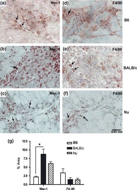
Immunohistochemistry for Mac-1 and F4/80 in the muscular lesion induced by bupivacaine (4 dpi) in C57BL/6 (a, d), BALB/c (b, e) and BALB/cnu/nu (c, f). The arrows show the positive cells. (e) Quantitative analysis of percentage of areas with positive immunolabelling for Mac-1 and F4/80 in the lesion area. Results are expressed as mean (±SD). Three animals per group were included. *P < 0.05. B6 = C57BL/6, Nu = BALB/cnu/nu.