
Figure 4.
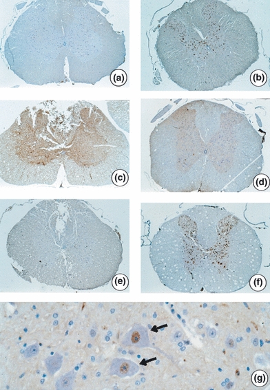
Photomicroscopy of spinal cord cross-sections adjacent to the epicentre of injury in Wistar rats. Immunohistochemistry with anti-NeuN. (a) Nuclei staining of intact neuronal cell bodies (NeuN-positive) in the grey matter in animal from GII (laminectomy only with placebo - 32 h) - 23.5×; (b) NeuN-positive neurons in the animals from GV (laminectomy only with placebo - 8 days) - 26×; (c) Mild staining of NeuN-positive neurons in animals from GI [spinal cord injury (SCI) with placebo - 32 h] - 24×; (d) Moderate staining of NeuN-positive neurons in animal from GIII (SCI with dantrolene - 32 h) – 21.8×; (e) Mild staining of NeuN-positive neurons in animal from GIV (SCI with placebo - 8 days) - 23.9×; (f) NeuN-positive neurons similar to the groups without injury. Animal VI (SCI with dantrolene - 8 days) - 23.3×; (g) Detail of NeuN-positive neurons (arrows) with nucleus staining by monoclonal anti-NeuN - 374.6×.