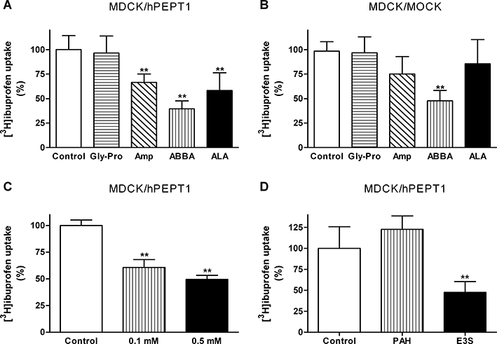
Figure 3.

Uptake of ibuprofen in Madin–Darby canine kidney cells (MDCK)/human proton-coupled peptide transporter (hPEPT1) or MDCK/Mock cells. Uptake was measured for 5 min with a buffer pH of 6.0. (A,B) Uptake of [3H]ibuprofen (0.25 µCi·well−1, 0.1 µM) in MDCK/hPEPT1 (A) or MDCK/Mock (B) cell monolayers in the absence or presence of 20 mM Gly-Pro, 20 mM ampicillin (Amp), 20 mM 2-(2-aminobenzoyl)benzoic acid (ABBA) or 20 mM δ-aminolevulinic acid (ALA). The results are mean ± SD of four to five individual cell monolayers. (C) Uptake of [3H]ibuprofen (0.25 µCi·well−1, 0.1 µM) in MDCK/hPEPT1 cells in the absence or presence of 0.1 mM or 0.5 mM ibuprofen. Results are mean ± SEM of 9–10 individual cell monolayers from three different cell passages. (D) Uptake of [3H]ibuprofen (0.25 µCi·well−1, 0.1 µM) in MDCK/hPEPT1 cells in the absence or presence of 1 mM p-aminohippuric acid (PAH) or 30 µM oestrone-3-sulfate (E3S). The results are mean ± SD of four individual cell monolayers. **P < 0.01, analysed by one-way anova followed by Dunnett's post-test.
