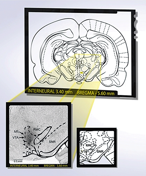
Figure 1.

Identification of the VTA as a microinjection site following post-mortem histological examination of microinjected Evans's blue. A rat was considered to be correctly injected when a black spot was seen in the VTA without any evidence of haemorrhage or necrosis. Diagram was modified from the atlas of Paxinos and Watson (1998). VTA, ventral tegmental area; SNR, substantia nigra reticular; ml, medial lemniscus; RMC, red nucleus magnocellular; MS, microinjection site. Scale bar: 0.5 mm.
