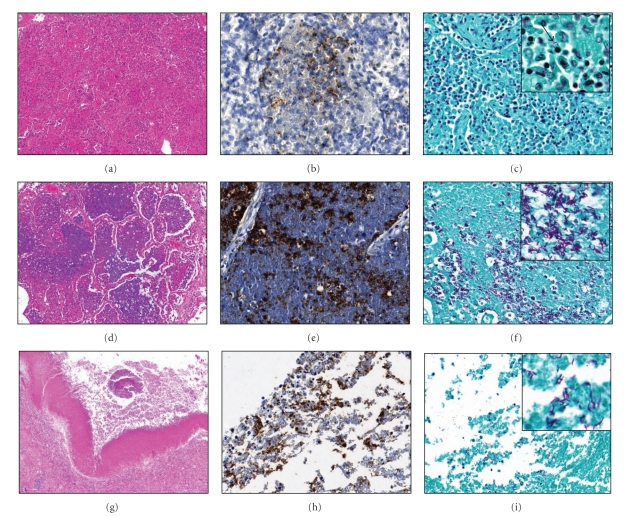
Figure 4.
Comparison of immunohistochemistry (IHC) for MTB and Zhiel-Neelsen acid fast staining. Early lipid pneumonia (a)–(c). (a) H&E stain demonstrates macrophages and lymphocytes within the alveoli, 100x. (b) MTB are noted on the IHC, 400x. (c) Few AFB are observed by Zhiel-Neelsen staining, 400x. Lipid pneumonia undergoing abrupt necrosis (d)–(f). (d) Cells in alveoli undergo necrosis and have a homogenous appearance, 100x. (e) Abundant mycobacterial antigens noted on the IHC stain for MTB, 400x. (f) Numerous acid-fast bacilli are observed, 400x. Cavitary TB (g)–(i). (g) H&E stain showing a cavity with a piece of detached lung and necrotic debris, 40x. (h) The wall of the cavity is strongly positive for MTB antigens, 400x. (i) Acid-fast bacilli are found lining the cavity wall, 400x.

