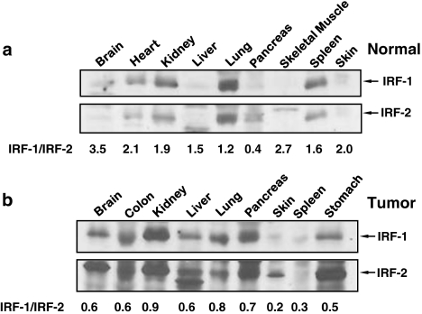
Figure 1.
Western analysis of IRF-1 and -2 expression in normal human and tumor tissues. Expression profile of IRF-1 and -2 in normal human tissues (a) or in human tumor samples (b) were analyzed by western immunoblotting (ProSci) and the IRF-1/IRF-2 ratio was quantified by densitometry using the Image J software

