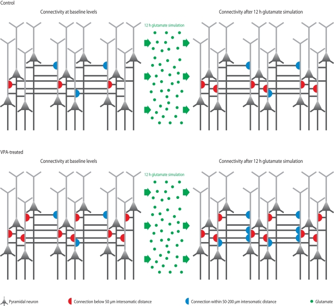
Figure 3.
Microcircuit hyper-plasticity in VPA-treated offspring. Depicted are schematic neural microcircuits from control (top) and VPA-treated offspring (bottom). In this experiment, brain slices were perfused for 12 h with a glutamate solution in order to stimulate the circuits and induce rewiring of connections (red and blue half-circles). The connectivity probability between neurons was accessed before (left panels) and after (right panels) the glutamate stimulation. In controls, the connection probability increased significantly within a range of less than 50 μm (red half-circles), which is the mini-columnar range, but did not change in ranges higher than 50 μm (measured up to 200 μm, blue half-circles), which is a columnar range. In VPA-treated offspring, the connections probability was already increased within the short mini-columnar range (below 50 μm, red half-circles) before the glutamate stimulation and did not increase any further after the stimulation (right panel), because the connection capacity was already boosted and probably saturated to a maximum at baseline levels. Indeed, controls only reached a similarly high connectivity probability within the mini-columnar range as VPA-treated offspring already exhibited at baseline levels after the 12 h stimulation was applied. However, in VPA-treated offspring, the connectivity probability increased significantly at ranges above 50 μm (right panel, blue half-circles), revealing a further remarkable rewiring capacity at the columnar range due to stimulation – a feature “normal” control microcircuits did not exhibit.

