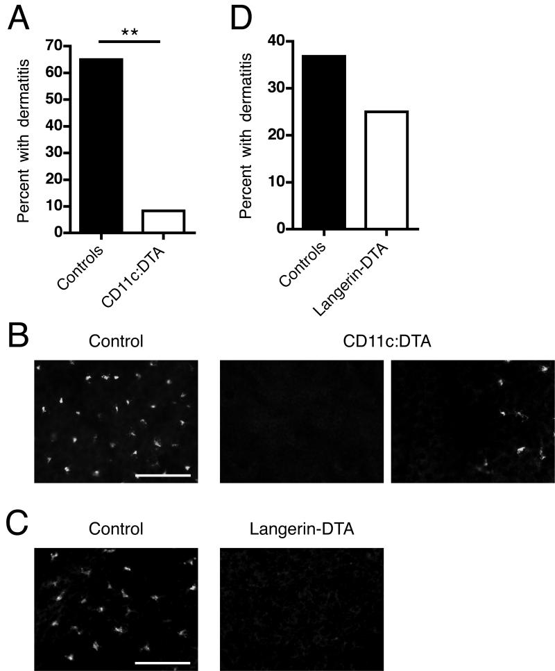
Figure 3. DCs other than LCs contribute to dermatitis.
(A) Percentage of female control (n = 20) and CD11c:DTA (n = 12) mice with dermatitis at 16 wk of age.
(B) Epidermal sheets from control and CD11c:DTA mice were stained for I-A/I-E (mouse MHC class II) to identify LCs (red). The images in the middle and on the right side represent 2 visual fields from the same sample to better illustrate that, rarely, small Langerhans cell clusters were detectable in CD11c:DTA mice. Data are representative of epidermal sheets from 3 mice of each type. Scale bar = 50 μm.
(C) Percentage of female control (n = 19) and Langerhans-DTA (n = 16) mice with dermatitis until death. The mean survival of Langerin-DTA mice was 145.2 days and of control mice 141.7 days.
D) Epidermal sheets from control and Langerin-DTA mice stained as in (B). Data are representative of epidermal sheets from 3 mice of each type. Scale bar = 50 μm. Statistics were calculated by Fisher’s exact test. **p < 0.01.

