Fig. 3.
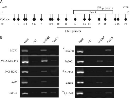
Histone modification status of MUC17 proximal promoter region in 10 cancer cell lines. (A) Schematic representation of the MUC17 gene promoter region. The relative positions of CpG sites and the ChIP primers used in this experiment are indicated. (B) ChIP analysis in the MUC17 promoter region was performed using antibodies against dimethylated H3-K9 (Me2-K9) and acetylated H3-K9 (Ace-K9). Input DNA was used as a positive control. NC indicates ChIP performed using rabbit IgGs as an isotype antibody control. Asterisks indicate MUC17-positive cell lines. Histone H3-K9 dimethylation was preferentially observed in cells with little or no MUC17 expression, whereas acetylation of histone H3-K9 was detected in all MUC17-positive cells. *MUC17-positive cell lines.

