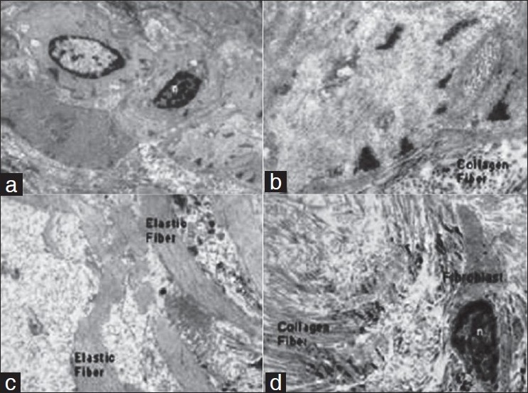
Figure 5.

Electron microphotograph of the deep fascia showing (a) Myofibroblast and nucleus (n) (× 4000). (b) Myofiberfilament with transverse section of collagen fibers adjoining to it (×4000). (c) Elastic fibers (× 4000). (d) Collagen fibers and fibroblast showing nucleus (×4000)
