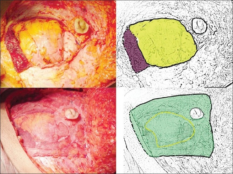
Figure 1.

Upper row: defect in the abdominal wall; the lower cut of rectus is depicted in brown and the defect in the wall in yellow in the drawing on left. Lower row: the use of Proline mesh. The mesh spans the defect and is sutured to the edge of defect, and contralateral anterior rectus sheath. The edge of the defect is depicted by broken yellow line and the mesh by green color in the drawing on left
