
Figure 2. Systemic Vascular Pathologies VEGFECKO mice.
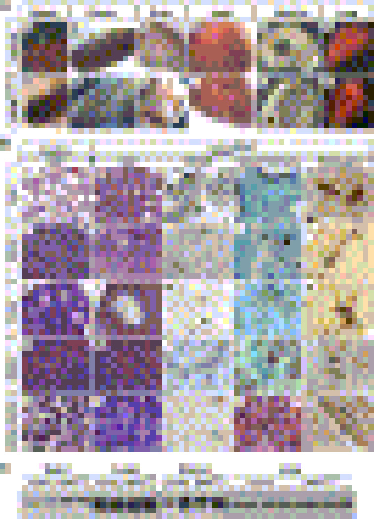
(A) Macroscopic analyses. a–f, Control. g–l, VEGFECKO. VEGFECKO organs of older mice show hemorrhage (arrows in g, i and l), tortuous and dilated vessels (arrows in h), areas of suggestive collagen accumulation in the heart indicative of microinfarcts (arrows, j) and intestinal perforations (arrows, k) (also see histology in Fig. S1).
(B) Histological sections of organs from younger mice. a–e, lung. Lung in VEGFECKO mice shows chronic inflammation (b), fibrosis (d) and excess of intravascular fibrin(ogen) deposits (e). PECAM staining reveals ruptured endothelial cells (arrowhead, c) and collapsed lumen (arrow, c). f–j, uterus. k–o, ovary. VEGFECKO ovary shows significantly enlarged vessels surrounding mature follicles (l) compared to wild type ovary (k). p–t, spleen. Asterisk indicates fibrosis. u–y, bone marrow. Fibrin(ogen) staining reveals clotting from VEGFECKO organs (arrows, e, o, t and y). Hemosiderin deposits were indicated by arrows in h, i, m, n, r, s, w and x. Bar, 100µm.
(C) VEGFR2 protein levels from VEGFECKO and control at 25 weeks were determined by immunoblots (same amount of protein was loaded per well). Slight differences in the uterus correlate with estrous cycle.