
Figure 6. Endothelial VEGF Mediates Survival in a Cell-Autonomous Manner.
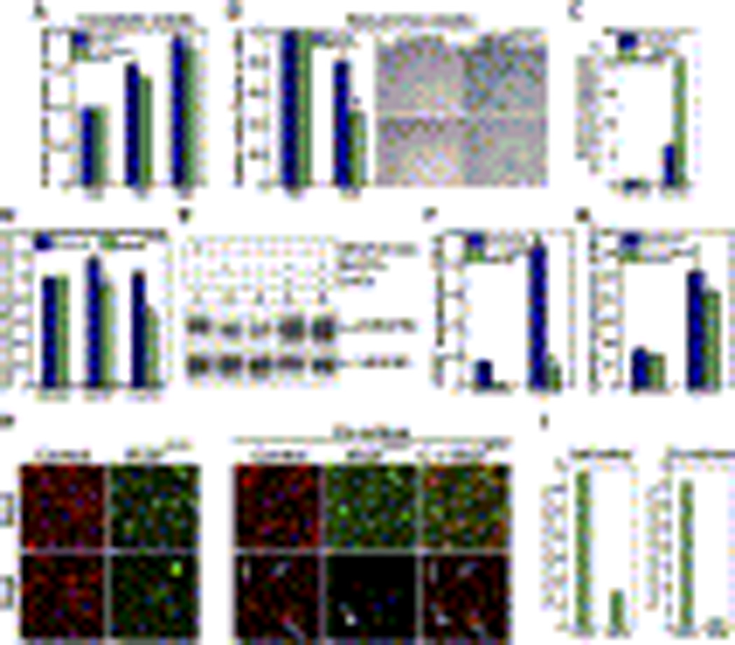
(A–B) Endothelial cells from control (blue) and VEGFECKO (green) mice were cultured in the presence (A) or absence (B) of serum and viable cells were counted at indicated times. Endothelial cells were stained for x-gal and nuclear fast red (right panels, B). Note the difference in cell number between control and VEGFECKO cells.
(C) Endothelial cells from control (blue) and VEGFECKO (green) mice were cultured in the presence of CoCl2, active caspases were fluorescence-labeled and quantified by fluorometer.
(D) Endothelial cells from control (blue) and VEGFECKO (green) mice were cultured in the presence of recombinant hVEGF165 or CoCl2 and viable cells assessed after 48hs.
(E) HUVECs were cultured under hypoxia for 24 h and analyzed for VEGFR2 phosphorylation (pVEGFR2, top) and VEGFR2 total levels (bottom). Lanes: 1, VEGF (100 ng/ml) for 5 min; 2–3, normoxia in the presence (2) and absence (3) of sodium orthovanadate (Na3VO4) for 24 h; 4–5, CoCl2 (hypoxia mimetic) in the presence of Na3VO4 for 24 h.
(F–G) Endothelial cells from control (blue) and VEGFECKO (green) mice were cultured in the presence of CoCl2 (100 µM) for 24 h and transcript levels of VEGF (F) and GAPDH (G) were determined by real-time RT-PCR. Relative RNA units (RRU) were normalized to β-actin. Data shown are means ± SD.
(H) Endothelial cells differentially labeled (Cy3 for wild type and BODIFY green for VEGFECKO) were cultured alone (left) or combined (right). Photographs were taken after 2 days and 5 days. Arrows indicate control cells (red). Arrowhead points to VEGFECKO cells (green).
(I) Quantification of cell survival following co-culture. Fluorescence labeled cells were counted at indicated times. Data are presented as percentage to control.