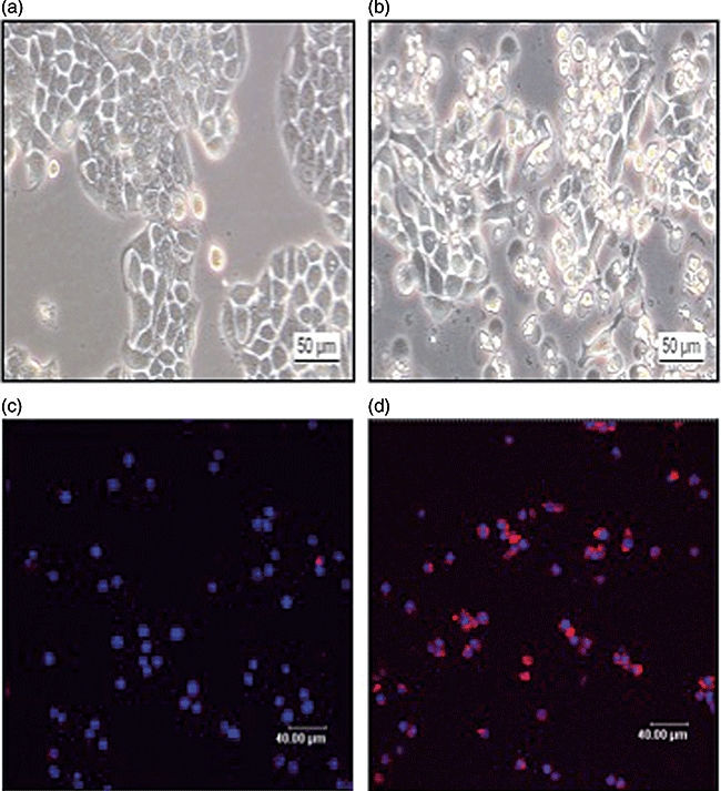
Fig. 3.

(a) Phase-contrast microscopy of native human colon epithelial cells (HT-29), which were cultured for 48 h in serum-free medium. (b) Phase-contrast microscopy of native HT-29 cells after stimulation with interferon (IFN)-γ (100 ng/ml). (c) Annexin staining of HT-29 cells cultured for 48 h in serum-free medium (coloured blue: staining of intact nuclei with Draq5). (d) Positive detection of externalized phosphatidylserine by annexin (coloured red) as a marker of apoptotic cells after treatment of HT-29 cells with IFN-γ (100 ng/ml) for 48 h.
