
Fig. 7.
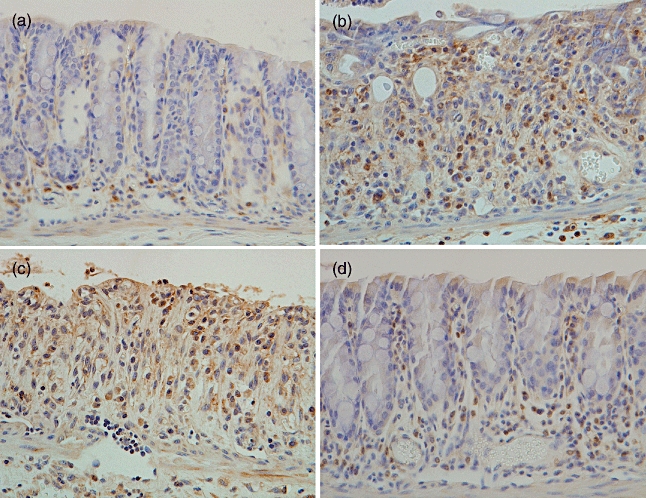
Immunohistochemical analysis for F4/80 in the colon. F4/80-positive staining was seen mainly in the mononuclear cells infiltrating the colon mucosa. (a) Non-treated mice. (b) Saline-treated mice given 3% dextran sulphate sodium (DSS) for 7 days. (c) pCAGGS-treated mice given 3% DSS for 7 days. (d) Migration inhibitory factor (MIF)/tetanus toxoid (TTX) DNA-vaccinated mice given 3% DSS for 7 days. Original magnification × 200. Representative pictures are shown. Similar appearances were observed in the colons of the other mice.