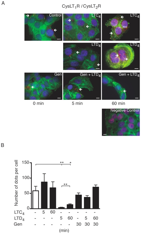
Figure 1. Receptor heterodimerization detected by a in situ proximity ligation assay (PLA).
(A) Briefly, Int 407 cells were grown to 50% confluency, stimulated with or without LTD4 (40 nM), LTC4 (40 nM), or pre-incubation with genistein (50 µg/ml) for 30 minutes. The receptor interactions were studied employing PLA, treated according to the manufacturer's instructions using the CysLT1R antibody (1∶250) and the CysLT2R antibody (1∶250) and mounted on glass slides with a fluorescence-mounting medium. Alternatively, the CysLT2R antibody was omitted as a negative control. The mounted slides were examined using a Nikon TE300 microscope (60×1.4 plan apochromat oil immersion objective), integrated in fluorescent microscopy. The detection of PLA-amplicons (red dots) was carried out using the “563 detection kit”. This kit includes the Hoechst 33342 dye for nuclear staining (blue) and the Alexa Fluor 488-phalloidin/actin for cytoplasmic staining (green). The red dots indicate close proximity between cellular bound antibodies, and they were counted using the MATLAB/Blobfinder software. (B) The data are given as percent of control and represent means ± S.E.M. of at least three separate experiments. The statistical analysis was performed with a Student's t test. *P<0.05 and ** P<0.01. The scale bar represents 10 µm.

