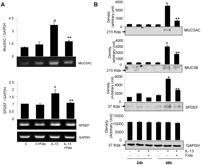
Figure 5. AR inhibition prevents IL-13-induced expression of Mucin and SPDEF in airway epithelial cell monolayer.
(A) The airway epithelial cell monolayer at ALI was serum starved without or with fidarestat and incubated with IL-13 for 18 h. Total RNA was isolated and subsequently RT-PCR was performed to assess the expression of Muc5AC and SPDEF. The bar diagrams show densitometric analysis of the corresponding blots (n = 4). *p<0.001 vs Control; **p<0.001 vs IL-13; (B) The monolayer of ciliated airway cells at ALI was treated with AR inhibitor for 24 h and subsequently incubated with IL-13 for 24 or 48 h. At the end of incubation, cell lysate was prepared and subjected to western blotting using antibodies against Muc5AC, Muc5B, and SPDEF. The membranes were stripped and re-probed with antibodies against GAPDH to show the equal loading of protein. A representative blot is shown (n = 4). *p<0.0001 vs Control; **p<0.001 vs IL-13.

