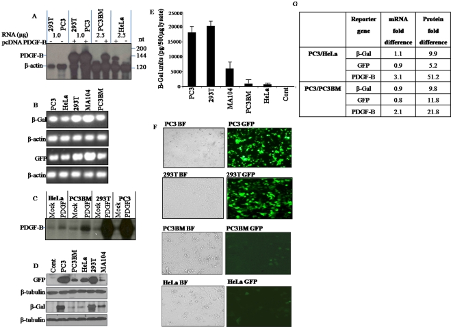
Figure 1. Analysis of RNA and protein levels derived from the transfected expression vectors of PDGF-B, GFP and β-Gal in PC3, PC3BM, HeLa, 293T and MA104 cells.
(A) PDGF-B mRNA levels were determined by RNase protection Assay. The 144 nt protected band corresponds to PDGF-B and the 120 nt band represents that of β-Actin mRNA. (B) RT-PCR of β-Gal, GFP and β-Actin mRNA in pcDNA3-β-Gal and pcDNA3-GFP transfected cells. (C) Radioimmunoprecipitation of PDGF-B protein expressed in pCMV-PDGF-B transfected cells using an N-terminal antibody [69]. (D) Levels of β-Gal, GFP and β-Tubulin proteins in pcDNA3- β-Gal and -GFP transfected cells. 50 µg of transfected cell lysate was analyzed for GFP and β-Gal levels by SDS-PAGE. (E) β-galactosidase assay using the β-Gal ELISA Kit from Roche Diagnostics. (F) Fluorescence microscopy and bright field (BF) images of 293T, PC3BM, HeLa and PC3 cells transfected with pcDNA-GFP reporter gene construct. (G) Analysis of the fold differences in expression of the reporter mRNA and protein levels between PC3 and HeLa, and PC3 and PC3BM.

