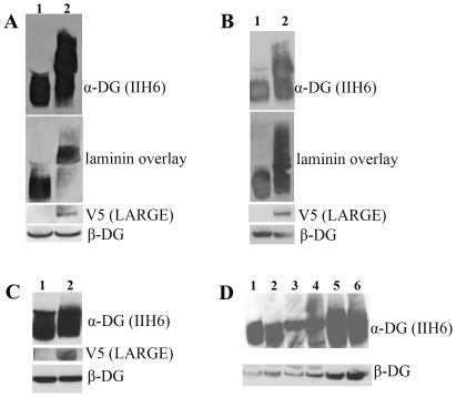
Figure 3. Western blot and laminin assay analysis of LARGE transgenic mouse tissues.
Western blot analysis of protein lysates from wild type and LARGE transgenic (line 68) tissues using antibodies to α-DG IIH6, β-DG, and V5. A. Wild type and LARGE transgenic quadriceps muscle. When the membrane was exposed for a short period using antibody IIH6 a clear band of higher molecular weight was detected in the samples from LARGE transgenic muscle compared to controls, while a longer exposure resulted in a continuous smear. Laminin-1 overlay showed that hyperglycosylated α-DG has increased laminin binding. β-DG expression was unaltered in transgenic mice. B. Wild type and LARGE transgenic mouse cardiac muscle. Laminin-1 overlay showed that hyperglycosylated α-DG bound laminin with a similar capacity to normally glycosylated dystroglycan. C. Wild type and and LARGE transgenic total brain lysates. No hyperglycosylated α-DG was seen using IIH6 in the LARGE transgenic brain samples even though the LARGE transgene (V5) was clearly expressed. D. Western blot analysis of protein lysates from control (1,3 and 5) and LARGE transgenic (2,4,and 6) mice derived from: small intestine (1,2), liver (3,4) and kidney (5,6). No hyperglycosylated α-DG was seen in any of these tissues. Expression of V5 (LARGE) was only detectable in heavily overexposed blots (data not shown). α-DG expression was unaltered in all tissue samples.

