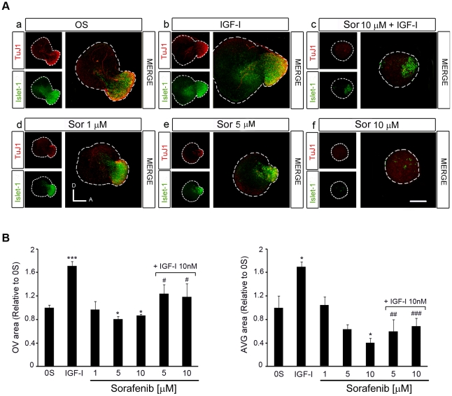
Figure 4. Inhibition of the RAF-MEK-ERK cascade impairs AVG formation.
(A) Otic vesicles were isolated from HH18 chicken embryos and incubated for 24 h in serum-free culture medium without additives (0S), with IGF-I (10 nM), Sorafenib, (Sor;1, 5 or 10 µM) or a combination of Sor (10 µM) and IGF-I. Whole otic vesicles were then immunostained for the ganglion neuroblast nuclei marker Islet-1 (green) and for the marker of neural processes, TuJ1 (red). Fluorescence images were obtained from the compiled projections of confocal images of otic vesicles. Representative images of at least five to six otic vesicles per condition and from at least three independent experiments are shown. Orientation: A, anterior; D, dorsal. Scale bar: 150 µm. (B) The otic vesicles (OV) and the acoustic-vestibular ganglia (AVG) areas were measured with Image Analysis Software (Olympus, Tokyo, Japan). The data are expressed as the mean ± SEM relative to the control value (0S) and they were compiled from the analysis of at least five to six otic vesicles per condition. Statistical significance was estimated with the Student's t-test: *P<0.05, ***P<0.005 versus 0S; #P<0.05, ##P<0.01 and ###P<0.005 versus IGF-I.

