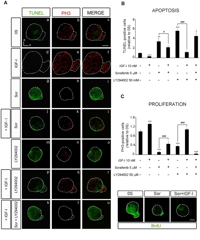
Figure 5. IGF-I partially rescues the effects of inhibiting RAF activity through the PI3K/Akt kinase pathway.
(A) Apoptotic cell death was visualized by TUNEL (green) in cultured otic vesicles and proliferation was detected with the mitosis marker Phospho-Histone 3 (PH3, red). Otic vesicles were isolated from HH18 chicken embryos and cultured for 24 h in serum-free medium without additives (0S, a–c), with IGF-I (10 nM, d–f), Sorafenib, Sor, (5 µM, g–i), LY294002 (50 µM, m–o), a combination of IGF-I and Sor (j–l), IGF-I and LY294002 (p–r) or IGF-I, Sor and LY294002 (s–u). (B) TUNEL positive or (C) proliferative PH3-labeled cells were quantified as described in at least 5 otic vesicles per condition. The results are shown as the mean ± SEM relative to the 0S condition. Statistical significance was estimated with the Student's t-test: *P<0.05, ***P<0.005 versus 0S; #P<0.05 and ###P<0.005 versus the indicated inhibitors; ∧P<0.05, ∧∧∧P<0.005 versus Sorafenib +IGF-I. Lower panels in C show BrdU (green) incorporation into cultured otic vesicles incubated for 24 h in the following conditions: 0S, with Sor (5 µM), or a combination of Sor and IGF-I (10 nM). Compiled projections of confocal images from otic vesicles are shown, and are representative of at least five to six otic vesicles per condition from three different experiments. Scale bar, 150 µm.

