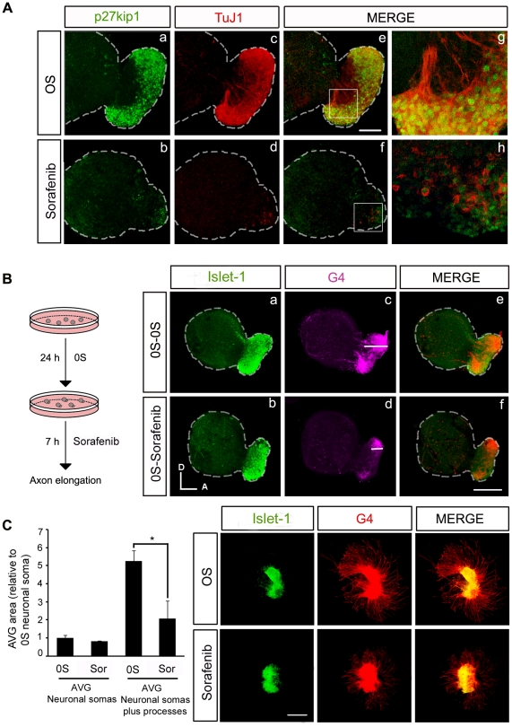
Figure 7. RAF kinase activity is required for the correct outgrowth of sensory otic neuron processes.
(A) Otic vesicles were isolated from HH18 chicken embryos and incubated for 24 h either in serum-free medium without additives (0S, a,c,e,g) or in the presence of Sorafenib (2.5 µM) (b,d,f,h). Immunohistochemistry of whole otic vesicles was carried out by double-staining for the nuclear cyclin-dependent kinase inhibitor p27kip1 (green) and for the marker of neural processes, TuJ1 (red). The boxed areas in panels e and f, correspond to the enlarged images in panels g and h respectively. Scale bar: 75 µm. (B) Otic vesicles were isolated from HH18 chicken embryos and incubated for 24 h in serum-free medium as in the 0S condition in A, and they were then incubated for a further 7 h without additives (0S-0S, a,c,e) or with Sorafenib (2.5 µM: 0S-Sorafenib, b,d,f). Whole otic vesicles were immunostained for the ganglion neuroblast nuclei marker, Islet-1 (green), and for the G4-glycoprotein marker of neuronal processes (G4, magenta). Note the differences in the magnitude of the white bars in the region in the acoustic-vestibular ganglia corresponding to the staining of neural processes in panels c and d. Scale bar: 150 µm. (C) Acoustic-vestibular ganglia (AVG) explants were obtained from stage HH19 chicken embryos and cultured in serum-free medium for 20 h with no additives (0S) or with Sorafenib (2.5 µM). Whole AVG explants were immunostained for G4 (red) and Islet-1 (green). Sorafenib-treated AVG have shorter processes. Scale bar: 300 µm. Fluorescence images were obtained from compiled projections of confocal images of otic vesicles and AVG. Bar graph on the left shows the quantification of the neuronal soma area in the AVG, which does not vary following Sorafenib treatment. In contrast, there is a statistically significant difference in the area of the AVG covered by processes (*P<0.05, Sorafenib versus 0S). Representative images of three independent experiments using five to six otic vesicles or AVG per condition are shown. Orientation: A, anterior; D, dorsal.

