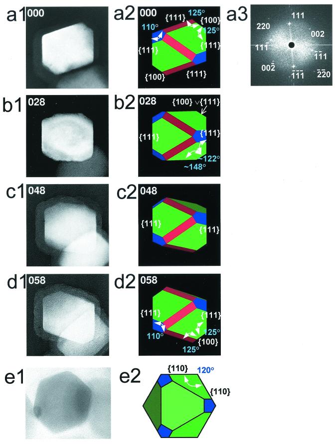
Figure 2.
Examples of a single truncated hexa-octahedral magnetite produced by magnetotactic bacteria strain MV-1 imaged with incremental stage rotation. Magnetites were extracted from cells and examined with a TEM at 200 kV. The degrees of rotation are noted in the upper left corner of each image. (a1) Magnetite, ≈35 nm in width, viewed at 0°. (a2) Simulated view of magnetite in a1. Intersecting {111} faces form a dihedral angle of ≈110° (theoretical angle is 109°); note these crystallographically equivalent faces are not equal in length. Intersecting {111} and {100} faces are at ≈125° (theoretical angle is 125°). Crystal is viewed down the [1–10] zone axis. (a3) The 2-D spatial Fourier transform calculated from the TEM image in a1. (b1) Same magnetite in a1 viewed at +28° from a1. (b2) Simulated view of magnetite in b1. {111} faces are bound by the edge defined by the intersection of {100} and {111} faces (represented by {100}∨{111}); this angle is ≈125° (theoretical angle is ≈122°). The intersection of {100}∨{111} edge and a {110} face is ≈145° (theoretical angle is ≈148°). (c1) Same magnetite in a1 viewed at +48° from a1. (c2) Simulated view of magnetite in c1. Position of the {111} faces indicates the magnetite is rotated about the long axis. (d1) Same magnetite in a1 viewed at +58° from a1. (d2) Simulated view of magnetite in d1. At ≈60° of rotation, the magnetite in a1 is now viewed down the [−101] zone axis displaying the 3-fold symmetry of this crystal. Intersecting {111} faces form an angle of ≈110°; again, note these crystallographically equivalent faces are not equal in length. Intersecting {111} and {100} faces are at ≈125°. (e1) Another magnetite, ≈40 nm in width, viewed down a [111] zone axis displaying a hexagonal projection. {110} faces intersect at ≈120°. (e2) Truncated hexa-octahedron in same orientation as magnetite in e1. Lengths of faces and angles are nearly identical to those of magnetite in e1.

