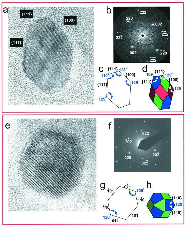
Figure 3.
(a) High-resolution TEM image of a single hexa-octahedral magnetite particle (≈35 × 25 nm) embedded in carbonate from Martian meteorite ALH84001. The carbonate matrix surrounding the magnetite is fine-grained, lacks long-range order, and shows no other recognizable structures (such as vesicular structure that would be expected by thermal decomposition of FeCO3 to Fe3O4). The crystal is aligned down the [1–10] zone axis and is elongated parallel to the [111] axis. (b) The 2-D spatial Fourier transform calculated from the TEM image shown in (a). (c) Outline of magnetite in a shows the {111} faces intersect at ≈110° (theoretical angle is 109°); {100} faces intersect {111} faces at ≈125° (theoretical angle is 125°). (d) The crystal habit we suggest for Martian magnetite (a) is the truncated hexa-octahedron that has a crystal habit identical to that described for biogenic magnetite crystals produced by strain MV-1 (see Fig. 2). The {100} blue faces are larger than those previously shown for the MV-1 magnetite consistent with variation in the degree of cubic faceting (also see Fig. 1b). None of the geometries of elongated biogenic magnetites in supplemental Fig. 4 adequately describes that of the ALH84001 magnetite in a. (e) High-resolution image of same crystal in a rotated 90° and viewed down the [111] zone axis. (f) Electron diffraction pattern of the TEM image shown in e. (g) Outline of magnetite in e shows the {110} faces intersect at ≈120° (theoretical angle is 120°). (h) Simulated truncated hexa-octahedron viewed down the [111] axis, same orientation as magnetite in e.

