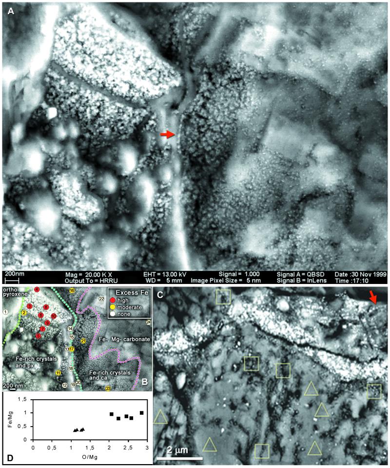
Figure 4.
(A–C) SEM-BSE micrographs of magnetite crystals in the rim region of carbonate globules in ALH84001. (A) Imaged through the intact surface of a freshly fractured specimen, chain of large elongated crystals (arrow), further chains of smaller crystals and other details in Fig. 1. (B) Energy-dispersive x-ray spectrometry analyses of elemental composition showing position of Fe-rich rim (detailed data are presented in Appendix 2, which is published as supplemental data on the PNAS web site, www.pnas.org). (C) Similar site in a resin-embedded, sectioned, and polished specimen, one chain marked by arrow, also site of Auger electron spectroscopy analyses. (D) Results of analyses of areas outlined in C: squares, magnetite crystal chains present; triangles, absent; ca, carbonate; px, orthopyroxene.

