
Figure 2.
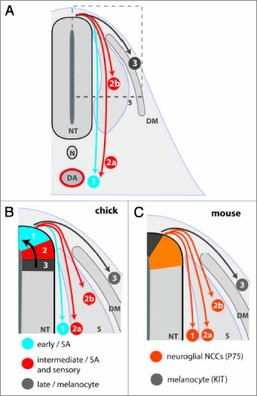
NCC derivatives are colonized in a ventral-dorsal pattern. (A) Schematic representation of a transverse section through the embryo trunk. NCC derivatives are colonized in a ventral to dorsal order. (1) The NCC precursors of the sympathetic ganglia emigrate from the neural tube first and migrate toward the dorsal aorta (DA). (2) The next wave of neural crest cells either migrate through the sclerotome to form sympathetic neurons (2a) or stall within the sclerotome to form the sensory ganglia (2b). (3) Melanocyte precursors emigrate last and scatter beneath the epidermis. (B and C) Schematic representation of the relationship between NCC migration and precursor localization in the neural tube. (B) Spatiotemporal fate map of NCC derivatives in the chick. NCC subpopulations emigrate successively from the neural tube, due to the progressive dorsal relocation of prespecified NCC precursors (black arrow). Thus, the dorsal tip is occupied by successive waves of sympathoadrenal, sensory and finally melanocyte precursors. The earliest wave of NCCs migrates to the dorsal aorta to seed the sympathetic nervous system (1). The intermediate wave of NCCs migrates through the sclerotome to form sympathetic neurons (2a) or stops within the sclerotome to form sensory neurons (2b). The final wave of NCCs migrates into the skin to form melnaocytes (3). (C) Relationship of NCC fate and precursor localization in the mouse neural tube. The KIT-expressing precursors of melanocytes are located at the dorsal tip of the neural tube throughout the period of NCC emigration, while the neuroglial precursors expressing p75 are located in a more ventral domain. The mechanism responsible for the ordered delamination of NCC subpopulations from the mouse neural tube has not yet been determined. The successive waves of NCCs detailed in (B) are represented numerically. NT, neural tube; DA, dorsal aorta; N, notochord; S, sclerotome; DM, dermomyotome.