
Figure 3.
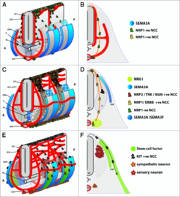
Schematic representation of NCC migration paths in the mouse. (A and B) The first wave of NCCs delaminates from the neural tube (NT) between E8.5 and 9.0 in the mouse and migrates towards the dorsal aorta (DA) to seed the sympathetic chain. (B) Half of a transverse section through the embryonic trunk shows the preferential migration of these NCCs in the intersomitic furrow alongside blood vessels (ISV). A small proportion of NCCs also migrate in the boundary between the anterior and posterior halves of the developing sclerotome (S). (C and D) The intermediate wave of NCCs delaminates between E9.0 and 10.5 in the mouse. These NCCs express NRP1 and are repelled by SEMA3A in the dermomyotome and posterior sclerotome and therefore migrate through the midst of the anterior sclerotome. ERBB2/3-expressing sympathoadrenal NCCs traverse the sclerotome to accumulate at the dorsal aorta in response to attractive NRG1 signals from surrounding mesenchyme. In contrast, sensory NCCs stall within the sclerotome, close to the neural tube. These cells likely express NRP2 in addition to NRP1. The NRP2 ligand SEMA3F is expressed in the posterior sclerotome to ensure that these NCCs remain in the anterior sclerotome. (D) Half of a transverse section through the anterior sclerotome shows the migration path of sympathoadrenal (yellow) and sensory (red) NCCs and highlights molecules that help to segregate their migration paths. (E and F) The late wave of NCCs delaminates between E10.5 and 14.5 in the mouse and gives rise to melanocytes. Stem cell factor in the dermomyotome (DM) and dermis attract KIT-expressing melanocyte precursors. By 10.5 dpc, sensory and sympathetic neurons have begun to differentiate to form the dorsal root ganglia and sympathetic ganglia. (F) Half of a transverse section through the anterior somite shows the characteristic migration path of melanocyte NCCs (grey) in relation to the position of the sensory and sympathetic ganglia. A, anterior; P, posterior; N, notochord. Blood vessels are shown in red.