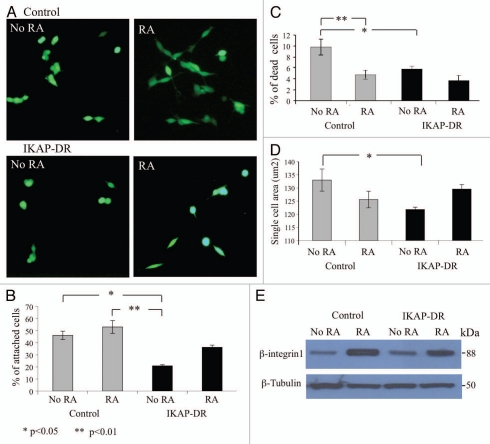
Figure 2.
Attachment and spreading assays. (A) Calcein stained control or IKAP-DR cells plated on laminin-coated plates without retinoic acid (NO RA) or with 10 µM retinoic acid (RA). (B) Graphic summary describing the averaged percentage of cells of three different experiments, that attached to the laminin coated plates in the indicated growth conditions. (C) Graphic summary of cell death indicated as percent of plated control or IKAP-DR cells. (D) Cell spreading described as the average cell area of a single cell of each strain in the indicated growth conditions. Statistical significance in (B–D) was determined using one-way ANOVA program, values are depicted at the bottom left corner of the figure. (E) Western blot analysis showing the expression of β-integrin1 in control and IKAP-DR cells grown on laminin without or with RA addition. Beta tubulin expression was used for normalization.

