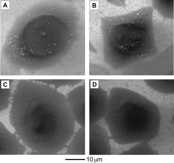
Figure 3.
(A, B) SEM images of SK-BR-3 cells after incubation with the PAA-coated (A) 45-nm Au nanospheres and (B) 33-nm Au nanocages at 37 °C for 24 h, respectively. (C, D) SEM images of SK-BR-3 cells after incubation with the PAA-coated (C) 45-nm Au nanospheres and (D) 33-nm Au nanocages at 37 °C for 24 h, respectively, followed by etching with 0.34 mM I2 for 5 min. The particle concentration of Au nanostructures in the culture medium was 0.02 nM.

