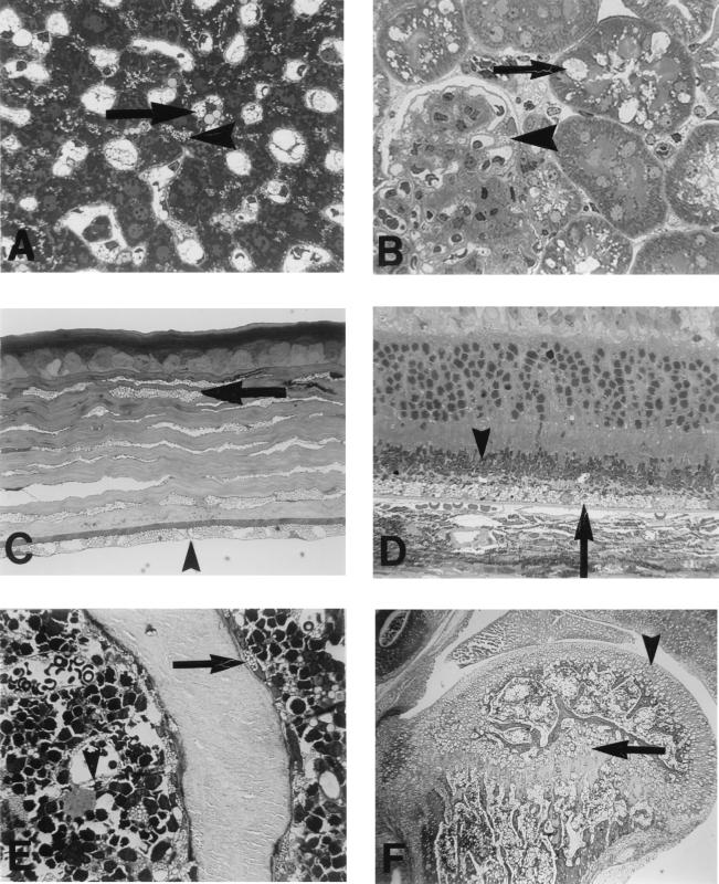
Figure 2.
Histopathology of the MPS VII/E540ATg mouse. (A) The liver from a 9-month-old MPS VII/E540ATg mouse has sinus-lining cells (arrow) distended by lysosomal storage. The hepatocytes also have a small amount of cytoplasmic vacuolization in a pericanalicular distribution (arrowhead) (Toluidine blue, 1 cm = 23.8 microns). (B) Renal tubular epithelial cells (arrow) in the kidney of a 9-month-old MPS VII/E540ATg mouse contained very large cytoplasmic vacuoles representing lysosomal storage. Glomerular visceral epithelial cells (arrowhead) and interstitial cells also have storage, although their cytoplasmic vacuoles are smaller than those seen in the tubular epithelial cells (Toluidine blue, 1 cm = 23.8 microns). (C) The cornea of a 3-month-old MPS VII/E540ATg mouse is altered with fibrocytes (arrow) and endothelial cells (arrowhead) distended with cytoplasmic vacuolization representing lysosomal storage (Toluidine blue, 1 cm = 23.8 microns). (D) Retinal pigment epithelial cells (arrow) from the eye of a 9-month-old MPS VII/E540ATg mouse have extensive storage and the outer segments of the photoreceptors (arrowhead) are disorganized (Toluidine blue, 1 cm = 23.8 microns). (E) The bone spicules from the rib of a 9-month-old MPS VII/E540ATg mouse are lined by osteoblasts (arrow) with cytoplasmic vacuolization. Similar lysosomal storage affects the bone marrow sinus-lining cells (arrowhead) (Toluidine blue, 1 cm = 23.8 microns). (F) A stifle joint from a 6-month-old MPS VII/E540ATg mouse has marked lysosomal storage in chondrocytes with distortion of the epiphyseal plate (arrow), articular cartilage (arrowhead), and periarticular articular connective tissue. The bone marrow also contains vacuolated cells (hematoxylin and eosin, 1 cm = 384 microns).

