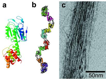
Figure 3.
Electron microscopy and model of a designed protein filament. (a) A ribbon model of a single molecule of the designed fusion protein. (b) A ribbon model of the protein filament as it was intended to assemble, with separate protein molecules colored differently. (c) Negatively stained electron micrograph of a bundle of filaments formed by the designed fusion protein. The bundle is 15–20 filaments across and reveals details indicative of the individual dimeric oligomerization domains that make up the fusion protein. In addition to bundles, networks of filaments were also observed in other micrographs.

