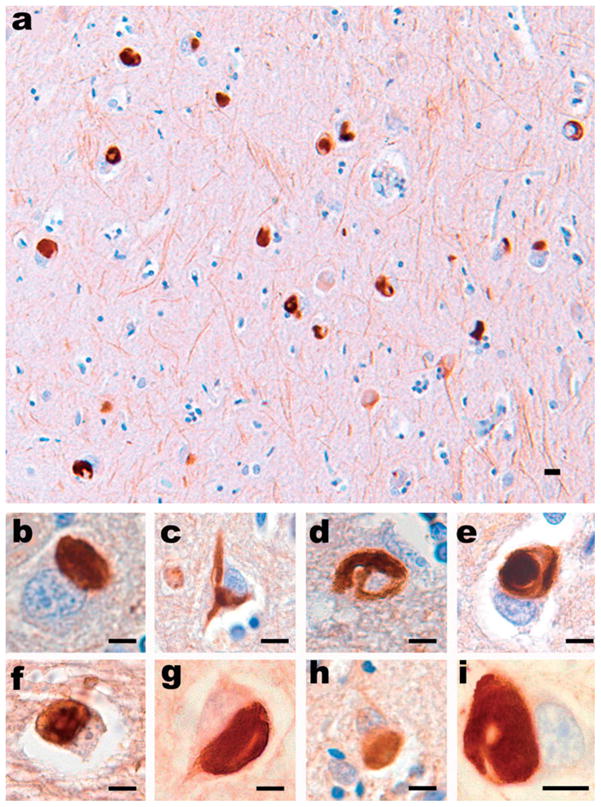
Figure 2.

All type IV neuronal IF proteins are present in the pathological inclusions of NIFID. (a) Neuronal inclusions in the subiculum of a case of NIFID contain α-internexin. α-Internexin immunohistochemistry. Neuronal inclusions in NIFID are pleomorphic. (b) Pick body-like inclusions are the most common morphological type. (c) A flame-shaped, NFT-like inclusion. (d) A filamentous serpiginous inclusion. (e) A globose NFT-like inclusion. α-Internexin immunohistochemistry. Epitopes of NF triplet proteins are present in inclusions of NIFID and are recognized by (f) phosphorylation-dependent NF-H; (g) non-phosphorylation-dependent NF-H; (h) phosphorylation-independent NF-M; and (i) phosphorylation-independent NF-L antibodies. NF immunohistochemistry. Scale bars = 10 μm
