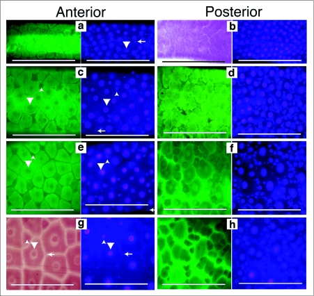
Figure 2.
Growth and development of the Culex pipiens larval midgut. Midguts from 1st through 4th instars were dissected, fixed and stained with Hematoxylin and DAPI. First through 3rd instar midguts were dissected approximately 12 hours after the molt. Fourth instar midguts were dissected 24 hours after the molt. Each subfigure has a left side image, taken using autofluorescence (green) or bright field (red), to emphasize the boundaries of the cells. The right side image is the same field of view to show DAPI stained nuclei. Large nuclei are indicated by downward pointing arrow heads. Intermediate sized nuclei are indicated by upward pointing arrow heads. Small oval nuclei are indicated by left pointing arrows. All images are taken at the same magnification and the scale bar represents 200 µm. (a) 1st instar anterior midgut have evenly spaced large nuclei with a few small nuclei between them, (b) 1st instar posterior midgut has evenly spaced large nuclei with more small nuclei between them, (c) 2nd instar anterior midgut has evenly spaced large nuclei and a few intermediate sized nuclei between them, (d) 2nd instar posterior midgut has evenly spaced large nuclei with many small and intermediate sized nuclei between them, (e) 3rd instar anterior midgut has evenly spaced large nuclei with a few intermediate sized nuclei between them, (f) 3rd instar posterior midgut has evenly spaced large nuclei with many intermediate and small sized nuclei between them, (g) 4th instar anterior midgut has evenly spaced large nuclei, a few intermediate sized and small nuclei between them, (h) 4th instar posterior midgut has many small and intermediate sized nuclei surrounding the large nuclei.

