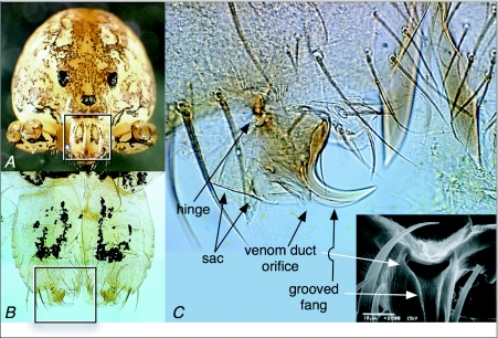
Figure 7.
External anatomy of the chelicerae and fangs of Scytodes thoracica. (A) Frontal view of S. thoracica. The legs and pedipalps of this specimen were removed so that they didn't obscure the face. The box outlines the region shown in B. (B) Anterior view of the chelicerae and fangs, both cleared with clove oil. The box outlines the region shown in C. (C) DICM (differential interference contrast microscopy) image of the fang and associated structures of a spitting spider after clearing with clove oil. Immediately above the hinge are two arrays of white, curved lines; these are the lyriform sense organs presumed to provide the spider with proprioceptive information about fang position. The DICM view is of the anterior surface of the distal portion of the spider's right chelicera, and thus shows the fang itself in side view. The inset, a scanning electron microscope image, gives an end-on view of the opening to the venom duct, with the sac above it and a groove leading from the orifice to near the tip of the fang.

