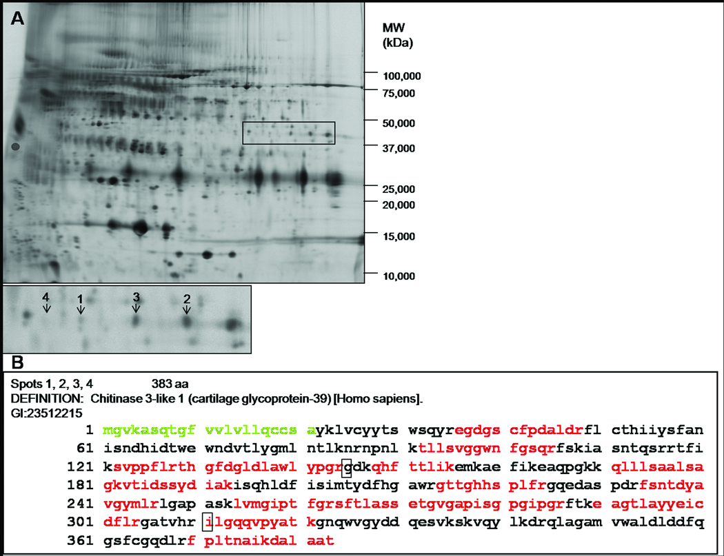
Figure 1.
(A) A representative 2-D DIGE image of CSF from the discovery cohort. Samples were depleted of six highly abundant proteins, fluorescently labeled, and subjected to isoelectric focusing followed by SDS-PAGE. YKL-40 is more abundant in four spots in the CDR 1 group (labeled 1–4 in the inset, with mean fold changes of 1.41, 1.50, 1.46, 1.32, respectively). The near invisibility of spot 4 in this printed representation illustrates the great sensitivity of 2-D DIGE to detect proteins of low abundance. (B) Sequence coverage of human YKL-40 by mass spectrometry. Indicated in red is the compilation of peptides identified in the four spots. The signal sequence is shown in green, and polymorphisms are indicated by boxes.

