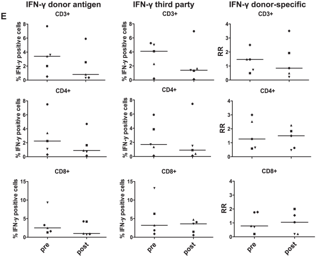
Figure 7. Comparison of IFN-γ production by T cells in CFSE-MLR before and 1 year after LTx.
CFSE-labeled PBMC from 5 LTx-patients were stimulated with donor-derived or 3rd party CD40-B cells, and re-stimulated with the same allo-antigens at day 5 of culture for 24 hours. During the last 15 hours Brefeldin A was added, and CFSE-dilution and intracellular IFN-γ was determined at day 6. Depicted are the percentages of T cells producing IFN-γ in response to donor-derived CD40-B cells and 3rd party-derived CD40-B cells, and the RR of IFN-γ producing T cells. No statistically significant differences were observed in comparing RR at 1 year after LTx versus before LTx (p≥0.44).

