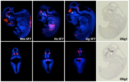
Figure 4. Optical projection tomography of 5F7-LacZ stained embryos.
Optical projection tomography (OPT) images of selected LacZ stained embryos. One representative embryo for each orthologous mouse human and chicken enhancer was selected. Arrowheads highlight expression in the trigeminal ganglion. Top: sagittal sections. Bottom: frontal sections. (Right) In situ hybridisations for Olig1 and Olig2 genes.

