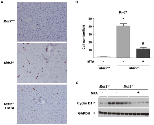
Figure 9. Increased hepatocellular proliferation in Mdr2−/− mice is attenuated by MTA administration.
Immunohistochemical staining of Ki-67 in liver sections from Mdr2+/+, control Mdr2−/−, and MTA-treated Mdr2−/− mice (Mdr2−/−+MTA)(A). Ki-67 positive hepatocytes were counted in 30 high-power fields per mouse, n = 5 per group (B). *P<0.01 vs Mdr2+/+ mice, #P<0.05 vs untreated Mdr2−/− mice. Western blot analysis of cyclin-D1 protein in liver extracts from Mdr2+/+ mice, untreated Mdr2− /− and MTA-treated Mdr2−/− mice (C).

