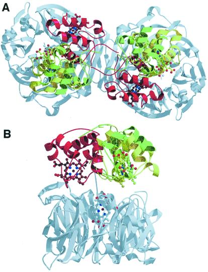
Figure 4.
Crystallographic structure of the two mutants of Pa-NiR. (A) Top view of the mutant proteins superimposed on the wt enzyme. Color code for the c-heme domains: dark green, H327A; light green, H369A; red, wt. It is evident that the c-heme domains glide away in a new configuration, which is almost the same for the two mutants. Notice also that the 3D structure of the d1-heme domain (gray) is identical for the three proteins. (B) Side view of the three structures (same color code); only one monomer is shown for the sake of clarity.

