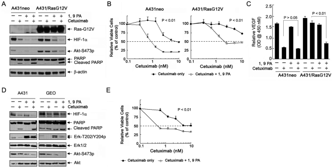
Figure 7. 1, 9 PA enhances responses of cancer cells expressing an oncogenic Ras mutant to cetuximab.
(A) Effect of 1, 9 PA and cetuximab, either alone or in combination, on the HIF-1α level and induction of apoptosis. A431neo and A431/RasG12V cells were untreated or treated with cetuximab (10 nM for 16 h), 10 µM 1, 9 PA (added the last hour before cell lysis), or both in 0.5% FBS culture medium. Cell lysates were prepared and analyzed by Western blotting with the antibodies shown. (B) 1, 9 PA-mediated sensitization to cetuximab-induced growth inhibition. A431neo and A431/RasG12V cells were treated with increasing concentrations of cetuximab ±5 µM 1, 9 PA in 0.5% FBS culture medium for 5 days. After treatment, the cells were subjected to an MTT assay. The optical density values of the treated groups were normalized to the values of the control groups (with or without 1, 9 PA treatment) and expressed as a percentage of respective control. The percentage of surviving cells was plotted as a function of treatment with increasing concentrations of cetuximab. The differences in cell survival between the two groups were statistically significant (p<0.01) when the concentrations of cetuximab were greater than 0.625 nM in A431neo cells and 1.25 nM in A431/RasG12V cells. (C) 1, 9 PA-mediated sensitization to cetuximab-induced inhibition of VEGF production. A431neo and A431/RasG12V cells were untreated or treated with 10 nM cetuximab, 10 µM 1, 9 PA, or both in 0.5% FBS culture medium for 16 h. The VEGF secreted into the conditioned media by the cells was measured by ELISA. The p-values for indicated comparisons were shown. (D) Comparison of A431 and GEO cells to treatment with 1, 9 PA and cetuximab, either alone or in combination. A431 and GEO cells were treated as described in (A). Cell lysates were prepared and analyzed by Western blotting with the antibodies shown. (E). 1, 9 PA-mediated sensitization GEO cells to cetuximab-induced growth inhibition. GEO cells were treated with increasing concentrations of cetuximab ±5 µM 1, 9 PA in 0.5% FBS culture medium for 5 days. After treatment, the cells were subjected to an MTT assay. The data were processed as described in (B). The differences in cell survival between the two groups were statistically significant (p<0.01) at all concentrations of cetuximab tested.

