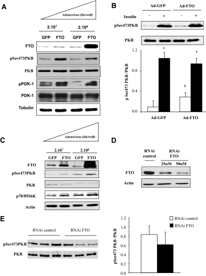
FIG. 3.
Adenoviral overexpression of FTO in differentiated myotubes. Human myotubes or C2C12 cells were infected with recombinant adenovirus encoding human FTO or GFP (control) for 48 h. A: Representative Western blots of FTO, pSer473PKB, PKB, pPDK1, PDK1, and tubulin, in GFP- or FTO-overexpressing C2C12 myotubes. B: Representative Western blots of pSer473PKB and PKB total, in GFP- or FTO-overexpressing C2C12 myotubes (210 [7] ifu/well), under basal conditions or after insulin stimulation. Histogram represents means ± SEM (n = 4). *P < 0.05 versus basal situation, #P < 0.05 FTO versus GFP. C: Representative Western blots of FTO, pSer473PKB, PKB, p70/85S6K, and actin in human myotubes overexpressing either GFP or FTO (210 [7] ifu/well). D and E: Myotubes were transfected with siRNA control or specific for FTO for 48 h. D: Validation of FTO silencing in muscle cells. E: Representative Western blots of pSer473PKB and PKB in myotubes silencing for FTO (50 nmol/l of siRNA). Histogram illustrates the quantification and normalization of the phosphorylation of PKB in myotubes silenced for FTO. Values are means ± SEM (n = 3). ifu, inclusion-forming units.

