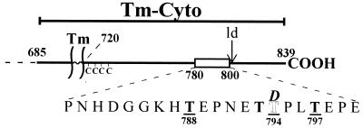
Figure 1.
Schematic drawing of the Tm-Cyto construct. The plasma membrane is represented by the pair of vertical lines. The 21-amino-acid region found to be important in N-CAM signaling is shown as an open box. The four threonines within this region are shown in bold. Underlined residues represent those whose substitution with alanine abolished the inhibitory activity of Tm-Cyto. Substitution of the threonine at position 794 (shown in outline) with aspartate (shown in italics) allows Tm-Cyto-mediated inhibition. The cytoplasmic cysteines (C) and the site of the ld insertion in the cytoplasmic domain are also indicated.

