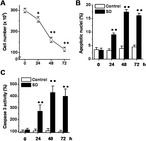
Figure 5.

Effects of serum deprivation (SD) on podocyte death. Cells were exposed to SD (24–72 h), and at the indicated time points, the cell number (A), the percentage of apoptotic nuclei (B) and the caspase 3 activity (C) were determined. Results are expressed as mean ± SEmean of four experiments run in triplicate. *P < 0.05; **P < 0.01 versus basal level; ••P < 0.01 versus control.
