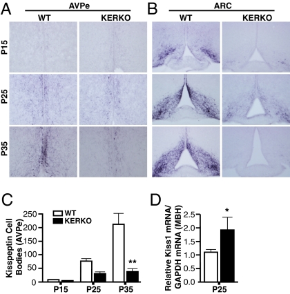
Fig. 3.
Kisspeptin expression fails to develop normally in AVPe of KERKO mice. (A and B) Representative examples of immunohistochemical labeling of kisspeptin cells in the AVPe (A) and ARC (B) of WT and KERKO mice at P15, P25, and P35. (C) Statistical analysis of kisspeptin immunoreactive (kisspeptin-IR) cell numbers reveals the progression of kisspeptin expression from undetectable at P15, through intermediate numbers at P25, and highest numbers at P35 in WT mice; by contrast, kisspeptin-IR cells appear significantly lower in numbers in P35 KERKO mice. (D) Kiss1 gene expression is increased in MBH tissues of P25 KERKO mice, compared with tissues obtained from a corresponding group of WT/ERαlox/wt mice. Data are presented as mean ± SEM. *P < 0.01; **P < 0.001.

