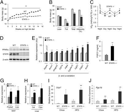
Fig. 3.
Resistance to dietary obesity and increased energy expenditure in STAT6-null mice. (A) Wild-type and STAT6−/− mice were placed on a HFD at 8 wk of age, and weight gain was monitored for 16 wk (n = 5 per genotype). (B) Dual-energy X-ray absorptiometry was used to determine body composition (n = 5 per genotype). (C) Indirect calorimetry was used to determine VO2 consumption in wild-type and STAT6−/− mice. (D) Immunoblotting for PPARα and STAT6 proteins in livers of wild-type and STAT6-null mice after 16 wk of HFD. (E) Quantitative RT-PCR analyses of total liver RNA from wild-type and STAT6−/− mice. Note induction of signature genes for β- and ω-oxidation in livers of STAT6−/− mice. (F) Serum Fgf21 levels were quantified by ELISA. (G and H) Quantitative RT-PCR analyses for adipose tissue and pancreatic lipases in WAT and liver of wild-type and STAT6−/− mice. Patatin-like phospholipase domain containing 2 (Pnpla2) and hormone sensitive lipase (Lipe) are expressed in WAT (G), whereas carboxyl ester lipase (Cel) and pancreatic colipase (Clps) are induced in liver (H). (I and J) Induction of uncoupled respiration in STAT6−/− WAT. Quantitative RT-PCR analyses for expression of Ucp1 (I) and Pgc1b (J) mRNAs in epididymal WAT of WT and STAT6−/− mice (n = 5 per genotype). Data presented as mean ± SEM. *P < 0.05; **P < 0.01.

