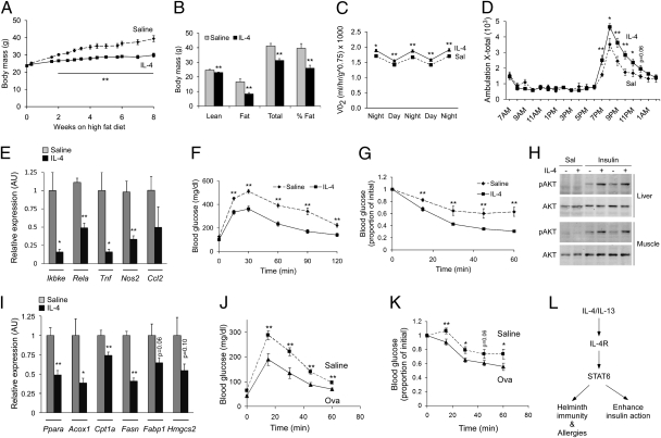
Fig. 5.
IL-4 and Th2 polarization improve glucose homeostasis in mice maintained on a HFD. (A and B) IL-4 treatment protects from diet-induced obesity. Total weight gain (A) and DEXA assessment of adiposity (B). Eight-week-old C57BL/6J mice (n = 6–8 per group) were placed on a HFD for 8 wk, and concurrently treated with saline (dashed line) or IL-4 (solid line). (C and D) Indirect calorimetry measurement of VO2 consumption (C) and locomotor activity (D) (n = 6 per treatment group). (E) Quantification of inflammatory genes in WAT of saline- or IL-4–treated mice by qRT-PCR (n = 6–8 per group). (F–H) IL-4 treatment improves glucose homeostasis. Glucose tolerance (F), insulin tolerance (G), and insulin signaling (H) was assessed in saline and IL-4–treated C57BL/6J mice (n = 6–8 per treatment group). (I) IL-4 treatment suppresses expression of PPARα and its target genes in liver. (J and K) OVA-induced Th2 polarization improves glucose tolerance (J) and insulin sensitivity (K) in D011.10 mice. Eight- to 10-wk-old D011.10 mice maintained on HFD were treated with saline (dashed line) or OVA with aluminum hydroxide (solid line) for 8 wk (n = 5 per group). (L) Model of IL-4/STAT6 functions in immunity, nutrient metabolism and longevity. Data presented as mean ± SEM. *P < 0.05; **P < 0.01.

