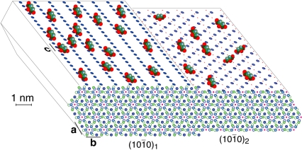
Fig. 5.
Schematic of apatite-bound citrate (with oxygen of the carboxylates in red) interacting with Ca2+ on two surfaces of high morphological importance of an idealized bone apatite nanocrystal, at a realistic citrate surface density of ca. 1/(2 nm)2. Calcium ions are blue filled circles on top and front surfaces, P is green (omitted on the top surfaces), OH- ions are pink dots, while phosphate oxygen is omitted for clarity. The hexagonal crystal structure projected along the c-axis (with greater depth of atoms indicated by lighter shading) shown in front reveals various layers of phosphate and calcium ions.

