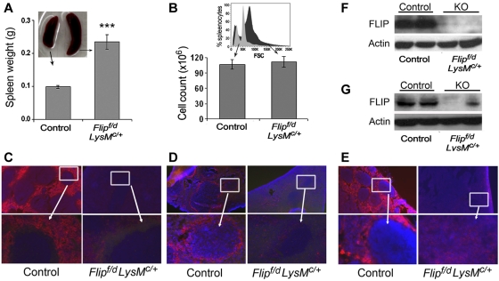
Figure 5.
Decreased macrophages and FLIP expression in the spleen and lymph node. Spleen size (n = 31; A) and total number of spleen cells (n = 16; B) in Flipf/d, LysMc/+ and littermate controls. A representative flow histogram of forward scatter (FSC) from ungated splenocytes of Flipf/d, LysMc/+ and littermate control mice is presented in the inset of panel B. Immunofluorescence microscopy of spleen was performed to identify red pulp macrophages (anti-F4/80; C) or marginal zone macrophages (anti-CD169; D), and of lymph nodes with anti-F4/80 antibodies (E). The data are representative of sections from 3-4 mice of each group. The area in the box is enlarged in the bottom panel. Randomly selected spleens (F) and lymph nodes (G) from littermate controls and Flipf/d, LysMc/+ mice were used to examine the expression of FLIP determined by immunoblot analysis. The data are representative of > 4 mice for each group.

