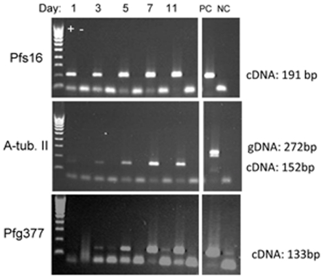
Figure 1. RT-PCR of sexual stage and sex specific proteins during gametocytogenesis.
Transcripts of Pfs16, α-tubulin II and Pfg377 were amplified from preparations of stage I–V gametocytes. Gel electrophoresis of amplified products; + and − refer to presence or absence of reverse-transcriptase in the cDNA reaction prior to amplification. PC: positive control; NC: negative control. Cultures were not 100% synchronous. All samples (including positive controls) were run on a single gel at the same time. Lower bands (<100 bp) in each panel, particularly prominent in the absence of cDNA amplification, are primer dimers. Minor contamination of pfg377 DNA is visible in lanes 4, 8 and NC, lower panel.

